We all have probably heard that “horses are fragile”, that “they simply break down easily”. But are those statements really true? Let’s look at an example of a disease that underlies over 60% of lameness cases and is the major cause for “loss of use” in horses 1.
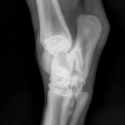
Osteoarthritis (OA) is a progressive disease of the joint 2. Degeneration of the articular cartilage, a pliable tissue located at the interface between the two bones, pain and joint dysfunction are the ultimate manifestations of OA. Once the pathological changes in the joint tissues begin, they are not likely to be reversed. At least, there is no clinical evidence that OA-associated pathology can resolve.
But why does OA develop in horses?
Unlike previously thought, OA is not a disease of normal “wear and tear”34. It is in fact a condition arising from the imbalance between the renewal and degradation of the joint tissues. Although OA is observed among the older individuals more frequently, horses of varying ages, including those that are very young, develop this degenerative condition as well.
The most consistent factor associated with the onset of this disease is overloading of the joints – forces that are too high in intensity and frequency for joint tissues to maintain their proper structure and function (read our previous article on Mechano-sensitivity, which explains how cells respond to mechanical stimulation)5. This does not mean that only horses exposed to high intensity training develop OA; horses involved in relatively low intensity work are also at risk, and oftentimes at a young age.
Why does OA occur in horses who lead a seemingly easy-going existence?
 The answer may lie in the fact that excessive forces acting on the joints can result not only from high intensity activities, such as jumping or racing, but also when a relatively low intensity repetitive force is acting on the joint in a wrong orientation or from a wrong direction.
What does this mean?
This means that if a horse’s joint experiences the maximum amount of loading or stress when it is not in its best alignment to manage this force, the damage can occur6,7. The reason being that the joint, and the entire musculoskeletal system for that matter, are designed to withstand the majority of stress through the reinforced structures arranged in a specific alignment. However, if the pressure/force comes from a slightly different direction or angle, the structure is simply unprepared to meet this stressor and suffers damage if this keeps repeating.
Knowing this, one can visualize that when the horse’s limb is not placed on the ground in the precise orientation it was designed for, especially if this happens repeatedly, like during trot or canter, the structures of the limb are likely to break down (read our article on Kinematics, which explains how small changes in movement patterns and forces affecting joints can be determined in horses).
What are the ways to prevent OA from occurring?
Based on what we discussed above - learning to recognize whether our horses’ movement patterns pose a risk to their joints and other limb tissues is an essential step in developing OA prevention strategies.
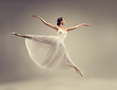
 Interestingly, in the field of human dance arts, ballet in particular, one of the directions towards injury prevention is to examine the entire body coordination in detail8. For instance, imbalance in back muscle function was found to be associated with ankle injuries. Why? Simply because movement coordination and placement of feet are not perfect when other structures in the body are asymmetric or are out of alignment. Similarly, scoliosis in humans, which results in an asymmetric alignment of the spine, also leads to asymmetric force distribution between the two feet during walking or running9.
In horses, imperfect spine alignment and dynamics during motion could also potentially lead to asymmetric stresses in limbs, perhaps explaining why one leg, and not both, display the pathology.
The prevailing thought in human rehabilitation medicine is that exercise and movement are essential for maintaining the joint functionality, even when OA has been already diagnosed. The innovation in human OA field lies in the design of individualized physical therapy strategies for each patient based on the exact location of their pathology and their specific movement patterns10. In fact, such an individualized approach towards designing physical activity regimens for patients is efficient and cost-effective in slowing down OA progression, and would improve the efficacy of the existing medical therapies.
To conclude
It is unlikely that our horses are designed to be fragile and to break down. However, our ability to recognize when structures are at risk of injury due to particular unfavorable movement patterns, and design of precision training strategies, are the key steps in preventing OA. These areas of research deserve equal attention as the development of therapeutic treatments for already existing conditions.
You can read our full review article about OA causes, treatments and new prevention strategies here.
References:
- McIlwraith, C. W., Frisbie, D. D. & Kawcak, C. E. The horse as a model of naturally occurring osteoarthritis. Bone Joint Res. 1, 297–309 (2012).
- Mcilwraith, C. W. & Acvs, D. From Arthroscopy to Gene Therapy-30 Years of Looking in Joints. (2005).
- Fu, K., Robbins, S. R. & McDougall, J. J. Osteoarthritis: the genesis of pain. Rheumatology 57, iv43–iv50 (2018).
- Yuan, X. L. et al. Bone–cartilage interface crosstalk in osteoarthritis: potential pathways and future therapeutic strategies. Osteoarthr. Cartil. 22, 1077–1089 (2014).
- DeFrate, L. E., Kim-Wang, S. Y., Englander, Z. A. & McNulty, A. L. Osteoarthritis year in review 2018: mechanics. Osteoarthr. Cartil. 27, 392–400 (2019).
- Cruz, A. M. & Hurtig, M. B. Multiple Pathways to Osteoarthritis and Articular Fractures: Is Subchondral Bone the Culprit? Vet. Clin. North Am. Equine Pract. 24, 101–116 (2008).
- Silver, F. H. & Bradica, G. Mechanobiology of cartilage: how do internal and external stresses affect mechanochemical transduction and elastic energy storage? Biomech. Model. Mechanobiol. 1, 219–38 (2002).
- Macintyre, J. & Joy, E. Foot and ankle injuries in dance. Clin. Sports Med. 19, 351–68 (2000).
- Daryabor, A. et al. Gait and energy consumption in adolescent idiopathic scoliosis: A literature review. Ann. Phys. Rehabil. Med. 60, 107–116 (2017).
- Skou, S. T. et al. Cost-effectiveness of 12 weeks of supervised treatment compared to written advice in patients with knee osteoarthritis: a secondary analysis of the 2-year outcome from a randomized trial. Osteoarthr. Cartil. (2020). doi:10.1016/j.joca.2020.03.009

